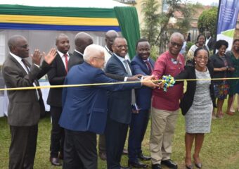
The Faculty of Medicine through the Mbarara University Medical Student’s Association (MBUMSA) together with the Ear Nose and Throat (ENT) department held a grand round on June 06, 2023 on the topic “Epistaxis”

The presentation and discussion were centered on the key features of Epistaxis the Epidemiology, Etiology, Pathogenesis, Evaluation and Management. Epistaxis also known as nosebleed being one of the most common ear, nose, and throat (ENT) emergencies that present to the emergency department or the primary care clinic and are two types of nosebleeds: anterior (more common), and posterior (less common, but more likely to require medical attention). The source of 90% of anterior nosebleeds is within Kiesselbach’s plexus (also known as Little’s area) on the anterior nasal septum. There are five named vessels whose terminal branches supply the nasal cavity:
1) Anterior ethmoidal artery, 2) Posterior ethmoidal artery, 3) Sphenopalatine artery, 4) Greater palatine artery, 5) Superior labial artery

Nosebleeds are rarely fatal, accounting for only four of the 2.4 million deaths in the United States. About 60% of people have experienced a nosebleed during their life, and only 10% of nosebleeds are severe enough to warrant treatment/medical intervention.
![]()
They occur most commonly in children ranging from 2 to 10 years old and the elderly ranging from 50 to 80 years old. There are multiple causes of epistaxis which can be divided into local, systemic, environmental, and medication-induced.

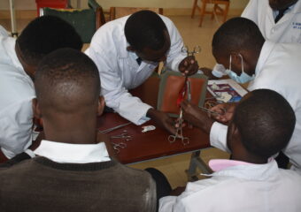
The students (Lugaaju Charles, Nyanzi Timothy Joseph, Mucho Caleb, Nsereko Joseph, Eric Ochen) discussed the basic anatomy and blood supply of the nose, common causes of nose bleeds, medicines that can predispose people to having a nosebleed, possible complications of nose bleeding and how to manage a patient that is bleeding from the nose. The grand round session was attended by medical students from different programs and years, both physically and online. In attendance also was Dr. Ruth Kagere, a lecturer and Ear, Nose and Throat specialist.
Local causes: 1. Digital manipulation, 2. Deviated septum 3. Trauma 4.Chronic nasal cannula use. Systemic causes: 1. Alcoholism 2. Hypertension 3.Vascular malformations 4. Coagulopathies (von Willebrand disease, hemophilia) Environmental factors: 1. Allergies 2. Environmental dryness (more common in winter months)
Medications:
- NSAIDs (ibuprofen, naproxen, aspirin)
- Anticoagulants (warfarin)
- Platelet aggregation inhibitors (clopidogrel)
- Topical nasal steroid sprays
- Supplement/alternative medications (vitamin E, ginkgo, ginseng)
- Illicit drugs (cocaine)
While epistaxis is a very common spontaneous problem, rarer etiologies such as neoplasms or vascular malformations must always be in the differential diagnosis, particularly if additional symptoms such as unilateral nasal obstruction, pain, or other cranial nerve deficits are noted.
 PATHOGENESIS :Nosebleeds are caused by the rupture of a blood vessel within the nasal mucosa. Rupture can be spontaneous, initiated by trauma, use of certain medications, and/or secondary to other comorbidities or malignancies. An increase in the patient’s blood pressure can increase the length of the episode. Anticoagulant medications, as well as clotting disorders, can also increase the bleeding time. Most nosebleeds occur in the anterior part of the nose (Kiesselbach’s plexus), and an etiologic vessel can usually be found on careful nasal examination.
These are often difficult to control and are associated with bleeding from both nostrils or into the nasopharynx, where it is swallowed or coughed up, presenting as hemoptysis. It can generate a greater flow of blood into the posterior pharynx and have a higher risk for airway compromise or aspiration due to increased difficulty in controlling the bleed.
 EVALUATION: Differentiating an anterior or posterior is key in management. Diagnosis of anterior bleeding is can be made by direct visualization using a nasal speculum and light source. A topical spray with anesthetic and epinephrine may be helpful for vasoconstriction to help control bleeding and to aid in the visualization of the source. Usually, the diagnosis of posterior bleeding is made after measures to control anterior bleeding have failed. Labs may be obtained if necessary, including a complete blood cell count (CBC), type and cross match, and coagulation studies, though should not delay treatment of an active bleed. Imagings such as x-ray or computed tomography have no role in the urgent or emergent management of active epistaxis.
MANAGEMENT AND TREATMENT
Start with a primary survey and address the airway, ensure the airway is patent. Next, assess for hemodynamic compromise. Obtain large-bore intravenous access in patients with severe bleeding and obtain labs. Reverse blood clotting as necessary, if there is a concern with medication use. The monitoring of oxygen and hemodynamic stability is vital.
 Treatment for anterior bleeding can be started with direct pressure for at least 10 minutes. Have the patient apply constant direct pressure by pinching the nose over the cartilaginous tip (instead of over the bony areas) for a few minutes to try to control the bleed. If that is ineffective, vasoconstrictors such as oxymetazoline or thrombogenic foams or gels can be employed. It is important to remove all clots with suction before any attempt at treatment is made. The reasons are twofold: 1) Clot will prevent any medication from reaching the vessel itself and 2) if packing becomes necessary, the clot can be pushed into the nasopharynx and aspirated. If topical treatments are unsuccessful, proceed with nasal examination to identify and cauterize the vessel with silver nitrate. If this too is unsuccessful, anterior nasal packing is necessary. This can be performed with absorbable packing material such as surgical or fibrillar, or with devices such as anterior epistaxis balloons, or nasal tampons (Rapid Rhino). If silver nitrate is used to cauterize a septal blood vessel, only use it on one side of the septum to prevent septal perforation. Thermal coagulation is painful and should rarely be attempted in an emergent setting.
Traditional petrolatum gauze can be used if one does not have access to balloons or tampons.
If all of these measures are unsuccessful, the patient should be intubated for airway protection and interventional radiology consulted emergently for embolization. If this service is unavailable, operative ligation of the sphenopalatine and ethmoid arteries can be performed in the operating room by an otolaryngologist.
 COMPILED BY NYANZI TIMOTHY JOSEPH